
Accurate diagnosis depends on precise imaging. Our Radiology department performs X-ray, mammography, CT, and MRI to support diagnosis; images are read by a physician certified as a diagnostic radiologist by the Japan Radiological Society.
X-ray

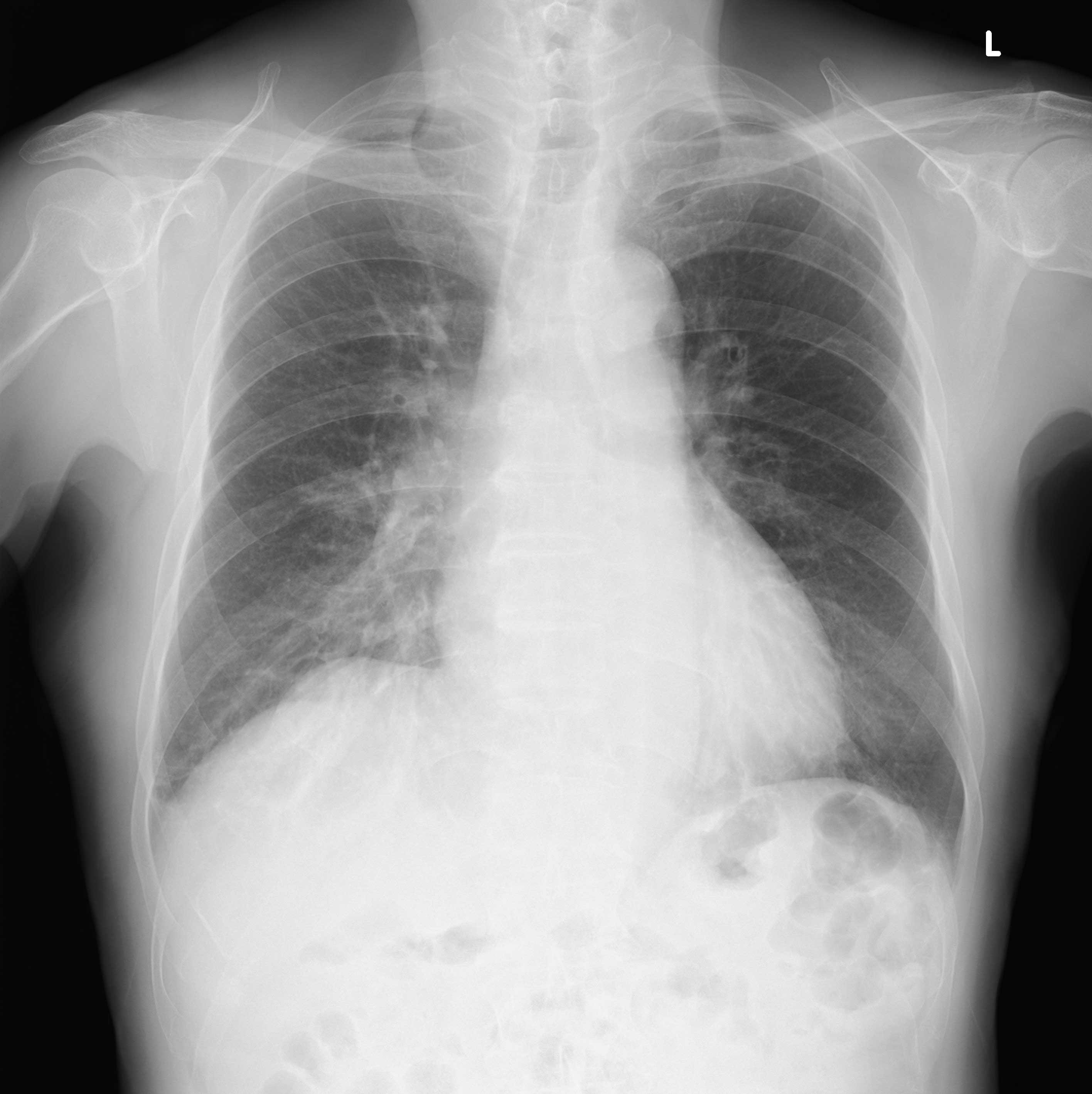
X-ray imaging of the chest, abdomen, bones, and joints. Please remove clothing with metal fasteners or buttons beforehand, since these can appear on the image.
Fluoroscopy
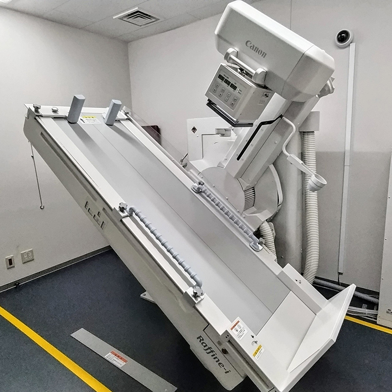
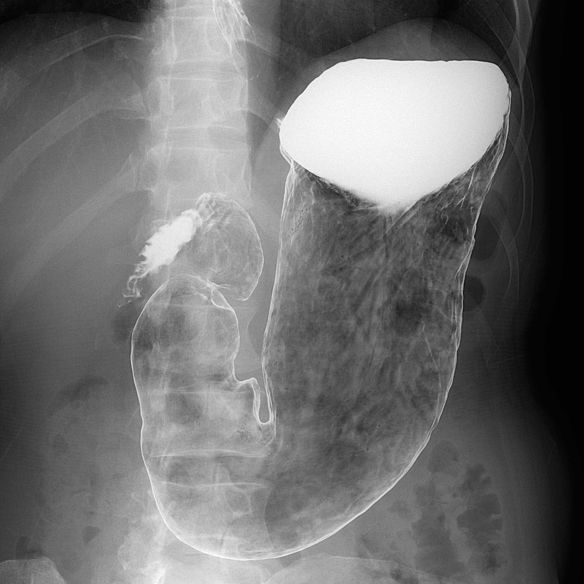
An upper gastrointestinal barium study images the oesophagus, stomach, and duodenum: you first drink a foaming agent to expand the stomach, then swallow barium so it coats the stomach lining, imaged under live fluoroscopy.
ERCP combines fluoroscopy and endoscopy to examine the gallbladder, bile duct, and pancreatic duct. Bronchoscopy likewise combines fluoroscopy and endoscopy to examine the lungs and airways. We also perform gastrostomy tube studies, bladder studies, and similar procedures.
CT
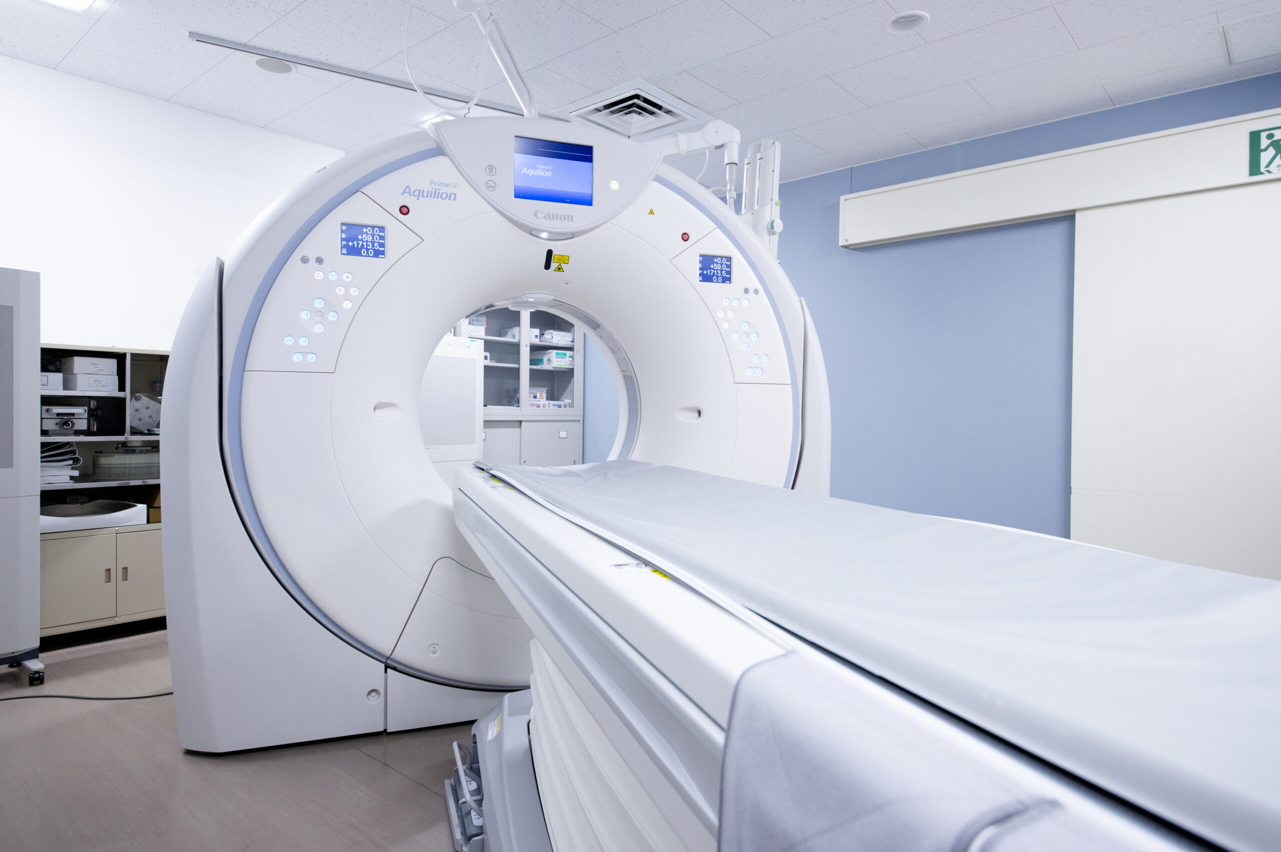
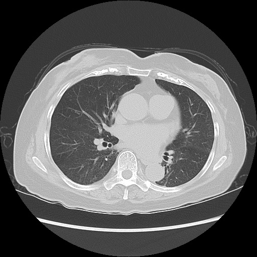
We use an 80-slice CT scanner, supporting lower-dose, high-quality images in a short scan (a breath-hold of just a few seconds). Scanning is tailored to what each department needs: in orthopaedics, for assessing joint calcification, tumours, and fracture; in health checkups, for visceral fat measurement supporting metabolic syndrome awareness, and low-dose lung cancer screening; and across internal medicine, surgery, and urology, for cancer and tumour assessment, with dose and technique adapted appropriately in paediatric imaging.
MRI
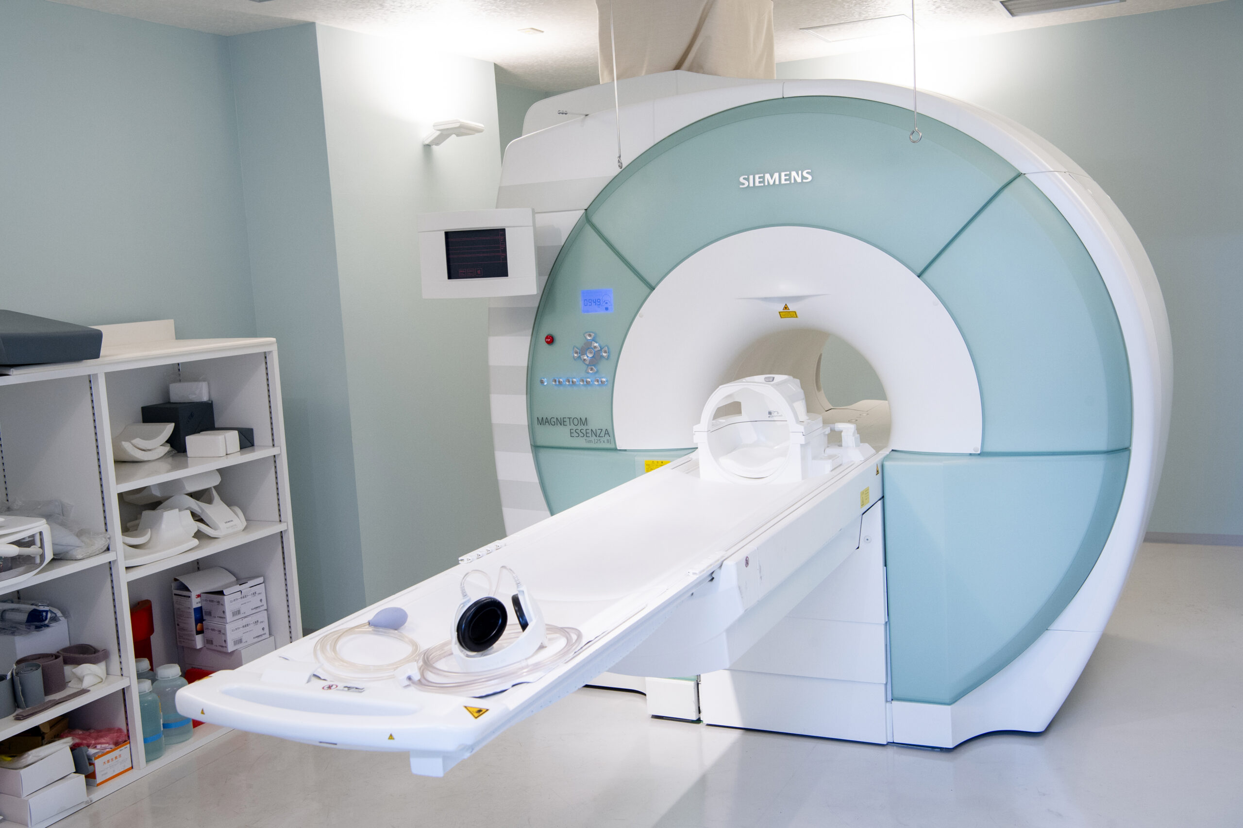
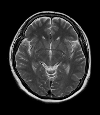
MRI uses a strong magnet and radio waves, rather than X-rays, to build a cross-sectional image of the body's interior from multiple angles.
MRI is less suited to a broad whole-body scan or to air-filled organs like the lungs, but is particularly good at showing detail in the brain, spinal cord, muscles and ligaments, and the uterus, ovaries, or prostate, areas that CT can find harder to resolve clearly. Because MRI does not use radiation, there is no radiation exposure to worry about, though a scan takes longer than CT (roughly 20 to 60 minutes depending on the type), can be noisy, and metal objects cannot be brought into the scan room, among other MRI-specific precautions. MRI may not be possible for some patients with claustrophobia or certain surgical history.
Our staff check for anything that would make MRI unsafe for you, both when your appointment is booked and again on the day, so your scan can proceed safely.
Mammography
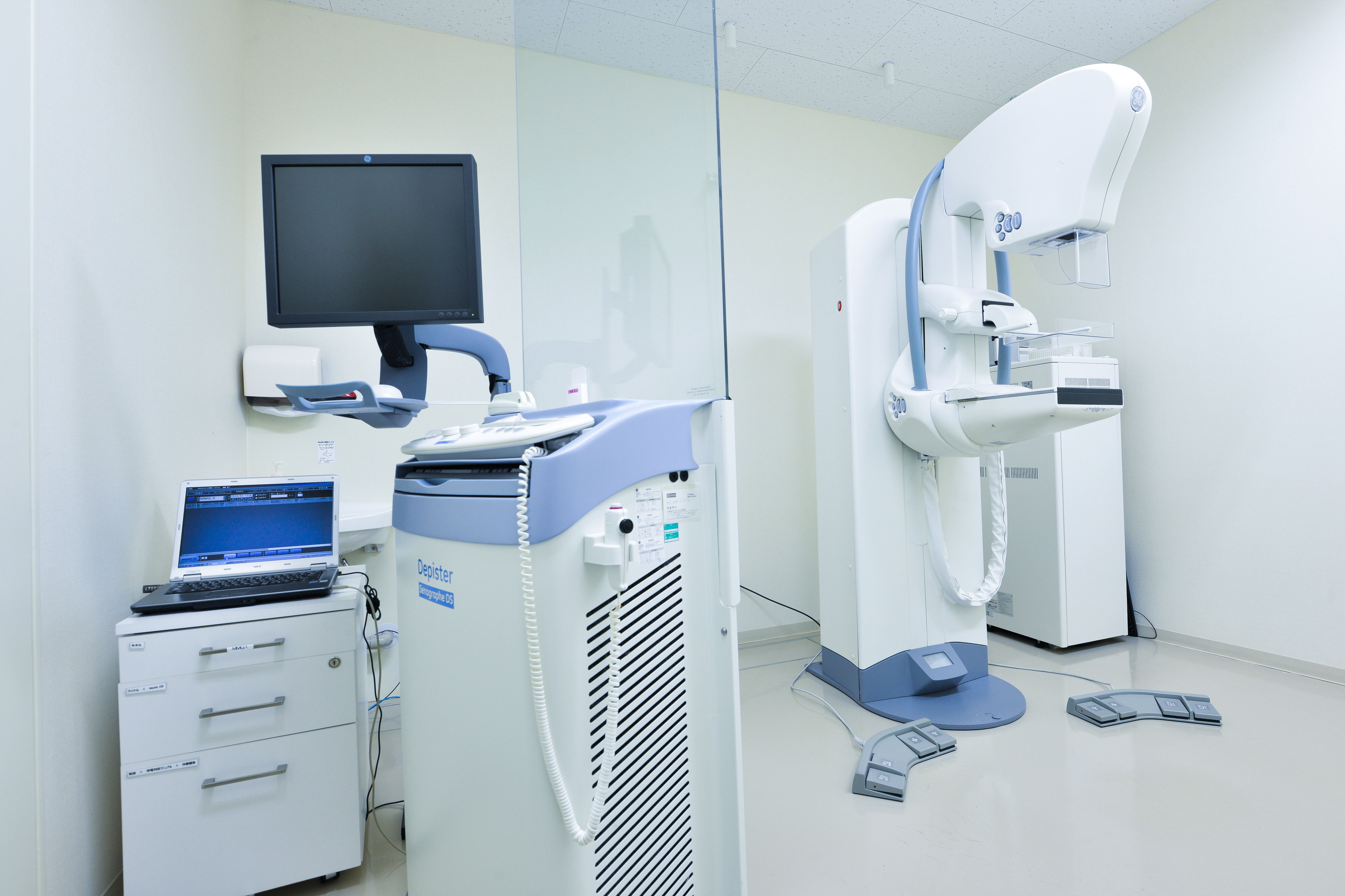
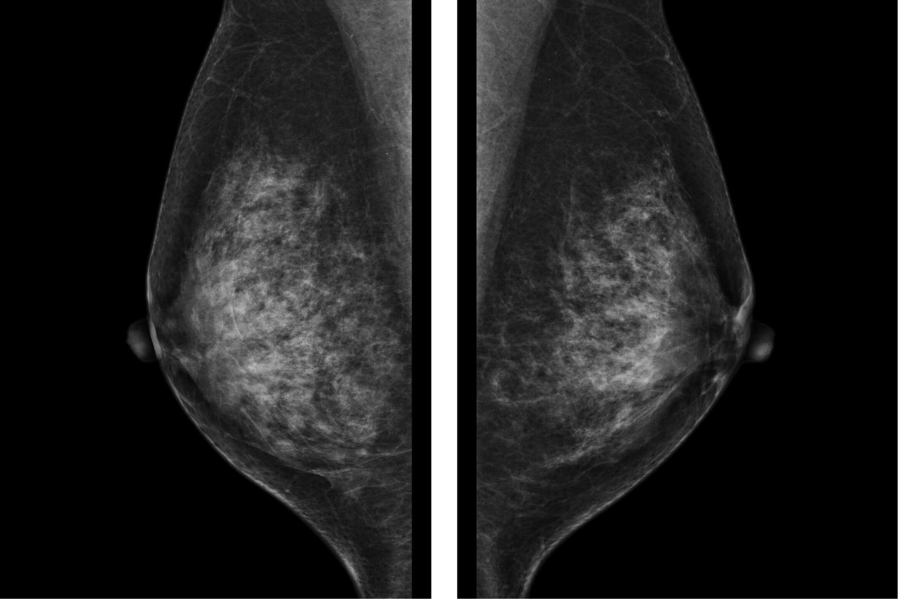
Mammography is an X-ray of the breast, able to detect a small breast cancer or a cancer that doesn't form a distinct lump, neither of which palpation alone would necessarily find. Because the breast is compressed between plates during the scan, some discomfort is possible, varying between individuals; this compression helps make breast tissue easier to see clearly, reduces radiation dose, and limits movement, all supporting a higher-quality image. We also offer mammotome biopsy to assess fine calcification visible only on mammography.
Mammography here is performed by a female radiologic technologist.
Bone density
We use DEXA scanning to assess bone density for osteoporosis diagnosis and monitoring. DEXA involves very low radiation exposure and gives a precise reading quickly; here, measurement is taken at the forearm (distal radius).
To book a visit, please use the form or call us. You can give up to three preferred dates, and choose from multiple time slots for each.